Home
/ Loculated Pleural Effusion Radiology - 1 - Conventional chest radiography and computed tomography (ct) scanning are the primary imaging modalities that are used for evaluation of all types of pleural disease, but ultrasound and magnetic resonance.
Loculated Pleural Effusion Radiology - 1 - Conventional chest radiography and computed tomography (ct) scanning are the primary imaging modalities that are used for evaluation of all types of pleural disease, but ultrasound and magnetic resonance.
Loculated Pleural Effusion Radiology - 1 - Conventional chest radiography and computed tomography (ct) scanning are the primary imaging modalities that are used for evaluation of all types of pleural disease, but ultrasound and magnetic resonance.. 8 kornecki a, sivan y. The formation of a transudate usually results from increased capillary hydrostatic pressure or from decreased colloid osmotic pressure. A prospective study of supine radiographs in 40 patients with pleural effusions showed that effusions with less than 175 ml are unlikely to be detected by this technique. Pleural fluid is seen extending to the right oblique fissure. Icu patients cannot sit up and the effusion layers posteriorly.
Air within a loculated pleural effusion is usually due to a bronchopleural fistula. Conventional chest radiography and computed tomography (ct) scanning are the primary imaging modalities that are used for evaluation of all types of pleural disease, but ultrasound and magnetic resonance. The parietal pericardium (arrow) clearly separates the loculated pericardial effusion (∗) from the pleural effusion (p). E7.8 loculated effusion loculated effusion. Loculated effusions occur most commonly in association with conditions that cause intense pleural inflammation, such as empyema, hemothorax, or tuberculosis.
Management Of Parapneumonic Pleural Effusion In Adults Archivos De Bronconeumologia from multimedia.elsevier.es In a study of 1,000 patients by proto ( , 2 ), the superomedial major fissure was seen in 8% of cases. On imaging, patients with entrapped lung have pleural effusions (which may be loculated), or an empyema. 9 stringel g, hartman ar. Sometimes in the setting of pleuritis, loculation of fluid may occur within the fissures or between the pleural layers (visceral and parietal). What are the different appearances of pleural effusion? The purpose of this study was to assess the value of intrapleural urokinase (uk) instillations in enhancing tube drainage of loculated, complex pleural effusions. However, effusion loculated in this part of the major fissure may mimic upper lobe disease because it manifests as increased opacity abutting the fissure line superiorly. 1 article features images from this case 20 public playlist include this case
Prior chest radiographs indicating that the blunting is a new finding also provide a good indicator of pleural effusion.
In chf effusions are bilateral and more on right. What are the different appearances of pleural effusion? A loculated effusion has an unusual shape (lentiform) or position in the thoracic cavity. Moreover, it is effective in guiding thoracentesis (thoracocentesis), even in small fluid collections 4. This creates a negative pressure environment in the pleural space, which fills up with fluid, creating a pleural effusion. Tube thoracostomy has variable success in the treatment of complex pleural effusions, with limitations because of viscous fluid, improper tube position or kinking, and, most. In the presence of pleural effusion, the elastic recoil of the lung causes each lobe to retract toward the hilum. Pleural effusions shift the mediastinal structures away from the side opacified. Contrary to the radiological method, ultrasound allows an easy differentiation of loculated pleural fluid and thickened pleura. A prospective study of supine radiographs in 40 patients with pleural effusions showed that effusions with less than 175 ml are unlikely to be detected by this technique. Loculated effusions are difficult to confirm with chest radiograph, but ultrasound, computed tomography (ct), and even magnetic resonance imaging (mri) may be used to verify a localized collection of pleural fluid. Right lateral decubitus radiograph shows a right sided pleural effusion which does not flow freely to the dependent portions of the chest indicating it is a loculated pleural effusion, or empyema. E7.8 loculated effusion loculated effusion.
This is the appearance of an empyema on a lateral decubitus chest radiograph. A prospective study of supine radiographs in 40 patients with pleural effusions showed that effusions with less than 175 ml are unlikely to be detected by this technique. On imaging, patients with entrapped lung have pleural effusions (which may be loculated), or an empyema. This is a loculated empyema. Note that the pigtail catheter in a is positioned below (rather than within) the loculated effusion, which explains why the catheter did drain any fluid until it was subsequently.
Malignant Pleural Disease European Journal Of Radiology from els-jbs-prod-cdn.jbs.elsevierhealth.com Normally, there is a similar retractile force applied to the entire pleural space by adjacent lung. On the pa radiograph, the loculated fluid collection manifests with incomplete borders, a radiographic sign of an extrapulmonary lesion, typically of pleural or chest wall origin. Pleural effusion is a common clinical finding with many potential causes 1 . Exudate ° ¸ ¸ 8. Prior chest radiographs indicating that the blunting is a new finding also provide a good indicator of pleural effusion. What are the different appearances of pleural effusion? The thickened visceral pleural peel may be visible on ct (figure 9). Other causes are complicated parapneumonic effusion , empyema, and tuberculosis.
A prospective study of supine radiographs in 40 patients with pleural effusions showed that effusions with less than 175 ml are unlikely to be detected by this technique.
When pleural malignancy is the underlying cause, pleural nodules or masses may be present. Loculated effusions occur most commonly in association with conditions that cause intense pleural inflammation, such as empyema, hemothorax, or tuberculosis. Contrary to the radiological method, ultrasound allows an easy differentiation of loculated pleural fluid and thickened pleura. More than one half of these massive pleural effusions are caused by malignancy; Discover (and save!) your own pins on pinterest Air within a loculated pleural effusion is usually due to a bronchopleural fistula. On the pa radiograph, the loculated fluid collection manifests with incomplete borders, a radiographic sign of an extrapulmonary lesion, typically of pleural or chest wall origin. Pleural effusion is an accumulation of fluid in the pleural space that is classified as transudate or exudate according to its composition and underlying pathophysiology. The purpose of this study was to assess the value of intrapleural urokinase (uk) instillations in enhancing tube drainage of loculated, complex pleural effusions. This is the appearance of an empyema on a lateral decubitus chest radiograph. Moreover, it is effective in guiding thoracentesis (thoracocentesis), even in small fluid collections 4. Pleural pseudotumor is a pleural fluid collection located within a lung fissure. Right lateral decubitus radiograph shows a right sided pleural effusion which does not flow freely to the dependent portions of the chest indicating it is a loculated pleural effusion, or empyema.
9 stringel g, hartman ar. Empyema is defined by purulent fluid collection in the pleural space, which is most commonly caused by pneumonia. The purpose of this study was to assess the value of intrapleural urokinase (uk) instillations in enhancing tube drainage of loculated, complex pleural effusions. The thickened visceral pleural peel may be visible on ct (figure 9). Moreover, it is effective in guiding thoracentesis (thoracocentesis), even in small fluid collections 4.
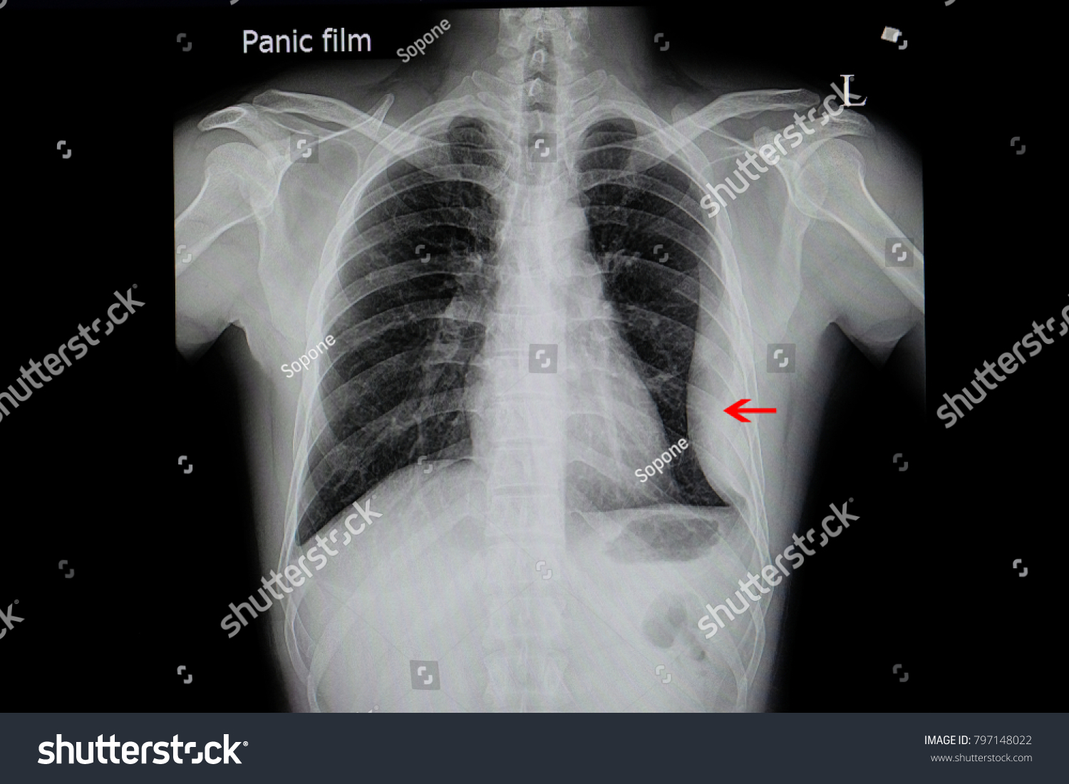
Chest Xray Film Patient Loculated Pleural Stock Photo Edit Now 797148022 from image.shutterstock.com Tube thoracostomy has variable success in the treatment of complex pleural effusions, with limitations because of viscous fluid, improper tube position or kinking, and, most. Intrapleural instillation of urokinase in the treatment of loculated pleural effusions in children. Moreover, it is effective in guiding thoracentesis (thoracocentesis), even in small fluid collections 4. Conventional chest radiography and computed tomography (ct) scanning are the primary imaging modalities that are used for evaluation of all types of pleural disease, but ultrasound and magnetic resonance. Loculation most commonly occurs with exudative fluid, blood and pus. Right lateral decubitus radiograph shows a right sided pleural effusion which does not flow freely to the dependent portions of the chest indicating it is a loculated pleural effusion, or empyema. Pleural effusions shift the mediastinal structures away from the side opacified. This is a loculated empyema.
E7.8 loculated effusion loculated effusion.
On imaging, patients with entrapped lung have pleural effusions (which may be loculated), or an empyema. Pleural pseudotumor is a pleural fluid collection located within a lung fissure. Treatment of loculated pleural effusion with intrapleural urokinase in children. Loculation most commonly occurs with exudative fluid, blood and pus. Air within a loculated pleural effusion is usually due to a bronchopleural fistula. Note that the pigtail catheter in a is positioned below (rather than within) the loculated effusion, which explains why the catheter did drain any fluid until it was subsequently. The formation of a transudate usually results from increased capillary hydrostatic pressure or from decreased colloid osmotic pressure. 31 long term drainage of an empyema may necessitate an eloesser flap, a surgically created fistula between the skin and pleural space. Pleural effusions shift the mediastinal structures away from the side opacified. 1 article features images from this case 20 public playlist include this case Normally, there is a similar retractile force applied to the entire pleural space by adjacent lung. Loculated effusions are difficult to confirm with chest radiograph, but ultrasound, computed tomography (ct), and even magnetic resonance imaging (mri) may be used to verify a localized collection of pleural fluid. Pa chest radiograph reveals a mediastinal mass, which is in continuity with the left heart border.
A loculated effusion has an unusual shape (lentiform) or position in the thoracic cavity loculated pleural effusion. Loculation most commonly occurs with exudative fluid, blood and pus.


 scanning are the primary imaging modalities that are used for evaluation of all types of pleural disease, but ultrasound and magnetic resonance.)